
In this guide
TECHNOLOGY, EXPLAINED VISUALLY
Equipment & illustrations
 Enlarge ↗
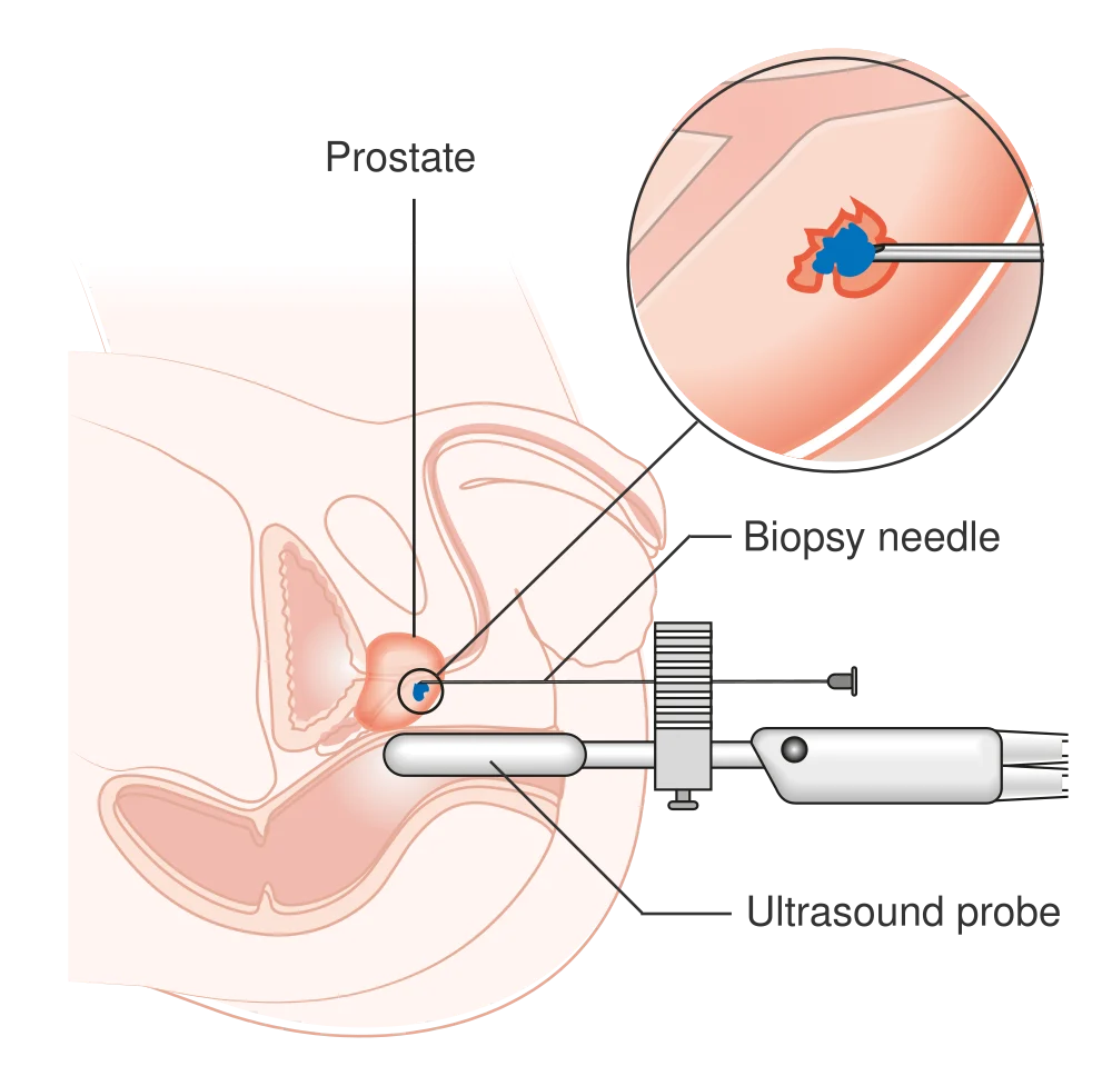
Transperineal biopsy
The biopsy needle reaches the prostate through the perineal skin. The ultrasound probe shown in the rectum guides the procedure.
Image: Cancer Research UK / Wikimedia Commons ↗ · CC BY-SA 4.0 · Resized and converted to WebP.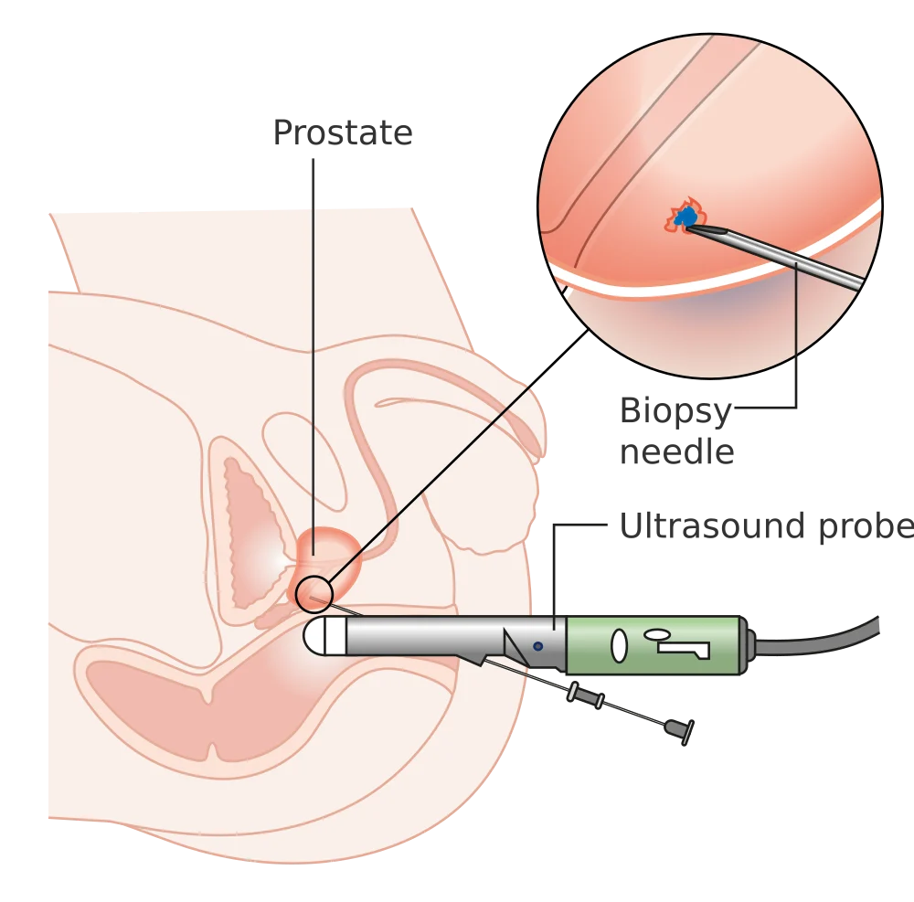 Enlarge ↗
Transrectal biopsy
The biopsy needle passes through the rectal wall into the prostate, with ultrasound guidance. The inset illustrates tissue sampling.
Image: Cancer Research UK / Wikimedia Commons ↗ · CC BY-SA 4.0 · Resized and converted to WebP.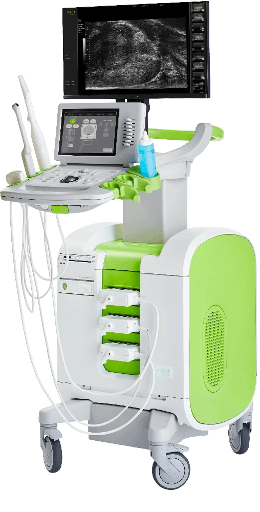 Enlarge ↗
ExactVu 29 MHz
High-frequency ultrasound provides detailed, real-time prostate imaging and can help guide targeted biopsy. Tissue analysis is still needed to diagnose cancer.
Image: Exact Imaging ↗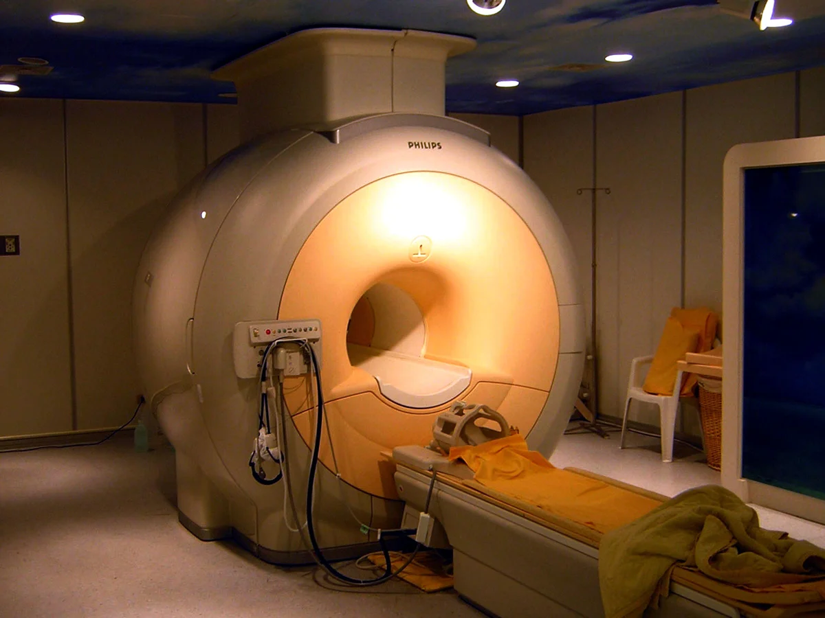 Enlarge ↗
Prostate MRI
MRI helps assess suspicious areas and local anatomy, often before biopsy. Shown: a Philips Achieva 3T MRI scanner, not a patient scan.
Image: KasugaHuang / Wikimedia Commons ↗ · CC BY-SA 3.0 · Resized and converted to WebP.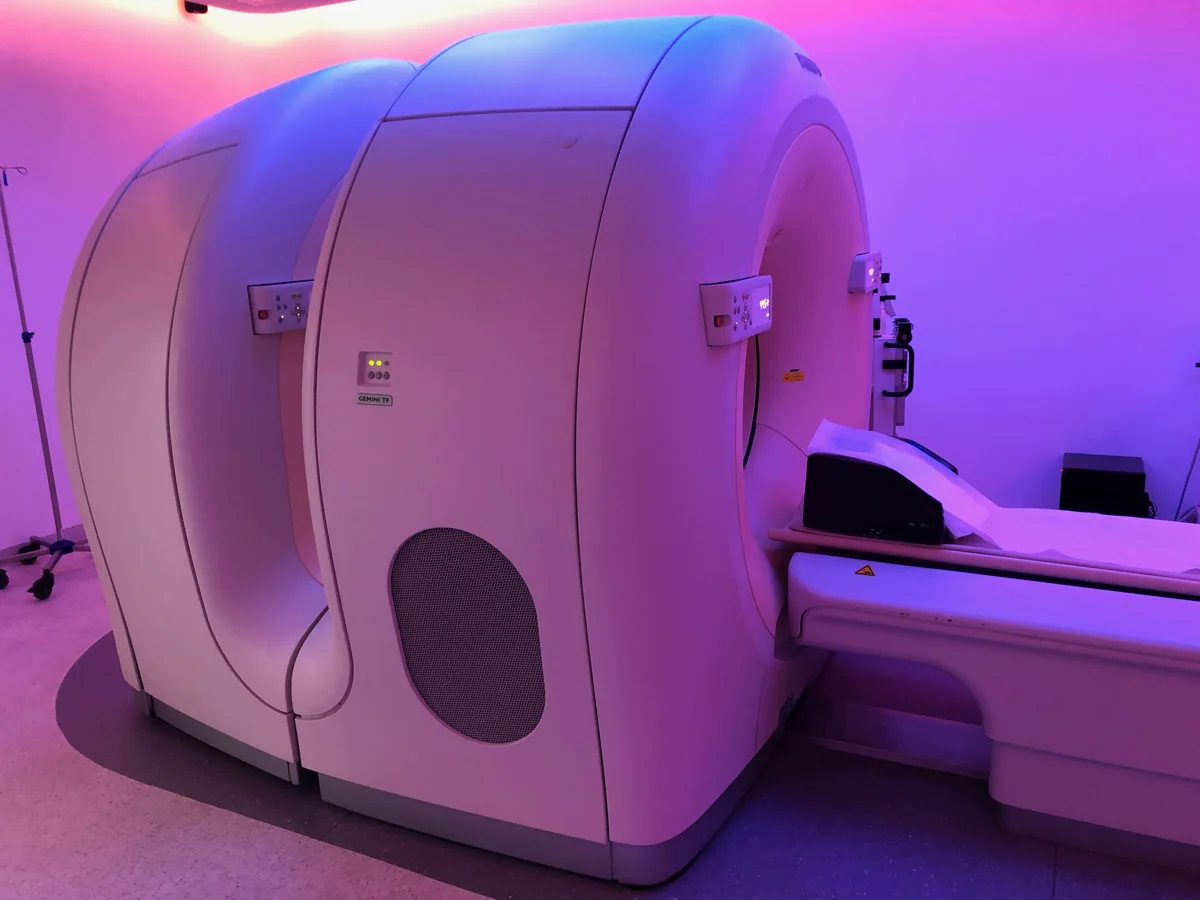 Enlarge ↗
PSMA PET/CT
PSMA PET/CT combines a PSMA-targeting radiotracer with PET and CT imaging. Shown: a generic Philips Gemini TF PET/CT scanner; the photograph is not a PSMA scan.
Image: Partynia / Wikimedia Commons ↗ · CC BY-SA 4.0 · Resized and converted to WebP.A stepwise diagnostic pathway
Not everyone needs every test. The aim is to identify clinically important disease while avoiding unnecessary procedures.
- Review the concernSymptoms, family history, previous PSA results, medicines and clinical examination.
- Refine the riskRepeat PSA when appropriate, interpret prostate volume and consider MRI or other imaging.
- Sample tissue when indicatedA targeted and/or systematic biopsy provides tissue for a pathologist to examine.
- Plan with the resultsUse grade, extent and appropriate staging to discuss monitoring or treatment.
PSAPSA testing & risk assessment
Prostate-specific antigen (PSA) is a blood marker used in prostate assessment and follow-up. A raised result does not by itself diagnose cancer. Results are interpreted alongside age, history, examination, prostate size and imaging.
Screening decisions · PSA trends · PSA densityTRUS & BIOPSYTransrectal ultrasound
TRUS uses an ultrasound probe placed in the rectum to image the prostate and assess its size. Ultrasound can also guide prostate biopsy; the needle may pass through the perineal skin or the rectum, depending on the planned approach.
TRUS imaging alone does not confirm or exclude prostate cancer. MRI, targeted biopsy and other findings are considered together.
Prostate imaging · Biopsy planning29 MHz MICRO-ULTRASOUNDReal-time prostate imaging
ExactVu 29 MHz micro-ultrasonography provides high-resolution, real-time views of prostate tissue to help identify areas for targeted biopsy. Its role is considered alongside MRI and the overall clinical assessment.
Micro-USG · Targeted biopsy · Transperineal biopsyGENOMICSTesting that can inform care
Inherited genetic testing and tumour genomic testing answer different questions. In selected patients, they can inform family risk or treatment choices. Tissue-based genomic classifiers may help refine risk when the result could change management.
Genetic counselling · Individual indicationsLearn more: NCI: PSA testing · ExactVu: 29 MHz imaging
Understanding each investigation
PSA: why a single number is not the whole answer
PSA can rise with benign enlargement, infection, inflammation or recent prostate procedures. Some medicines lower it. There is no single threshold that perfectly separates cancer from benign conditions; retesting and further assessment depend on context.
PSA density and free PSA
PSA density relates the PSA value to prostate volume. The free-to-total PSA ratio may add information in selected situations. These support risk assessment rather than replacing imaging or biopsy.
Prostate MRI and PI-RADS
MRI identifies suspicious areas and assists biopsy planning. PI-RADS expresses the likelihood of clinically significant cancer on imaging. A reassuring MRI reduces risk but cannot exclude all important cancers.
MRI fusion and targeted biopsy
MRI-visible targets can be matched to ultrasound during biopsy. Depending on the clinical situation, additional systematic or surrounding samples may be recommended.
Transperineal versus transrectal biopsy
Transperineal biopsy passes needles through the skin between the scrotum and anus; transrectal biopsy passes through the rectal wall. Ultrasound may be used with either route. Discuss anaesthesia, infection prevention, bleeding and temporary retention.
PSMA PET imaging
PSMA PET helps stage selected prostate cancers or investigate recurrence. It is not a routine replacement for MRI or biopsy in everyone with an elevated PSA.
Inherited testing, tumour testing and genomic classifiers
Inherited testing asks about genetic changes present throughout the body and may affect relatives. Tumour testing looks for changes that may guide treatment. Genomic classifiers estimate aspects of cancer behaviour; they are used selectively when they could alter a decision.
Further reading: NCI: PSA · EAU: diagnosis · NCI: biomarker testing
Prepare for your appointment
Bring the information that connects the picture.
- Dated PSA results and previous blood or urine tests.
- MRI and other scan images, as well as the written reports.
- Biopsy and pathology reports, if available.
- Your medicine list, including blood thinners and prostate medicines.
- Family cancer history, allergies and previous treatment details.
If a biopsy is planned, follow the specific preparation instructions. Do not stop blood thinners without an agreed plan.
Imaging guides the biopsy. Tissue establishes the diagnosis.
| Test | Main question | Important limit |
|---|---|---|
| PSA | Is further assessment appropriate, or is a known cancer changing? | A high PSA does not prove cancer; a low result does not exclude it. |
| MRI | Where are suspicious areas and what is the local extent? | Some important cancers can be missed. |
| TRUS | What is the prostate size and how can sampling be guided? | Routine ultrasound alone cannot rule cancer in or out. |
| 29 MHz micro-US | Are there suspicious real-time tissue patterns to target? | Operator experience and the overall diagnostic pathway matter. |
| Biopsy | Is cancer present in sampled tissue, and what is its Grade Group? | Sampling may not capture every focus; results must fit the whole assessment. |
| PSMA PET | Where might known cancer have spread or recurred? | Usually a staging or recurrence investigation, not a general screening test. |