
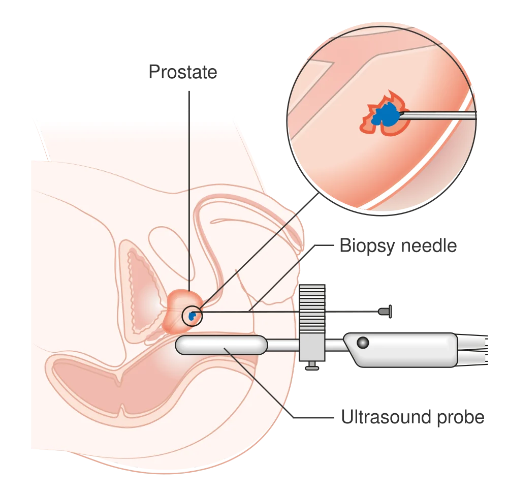
Transperineal
The needle passes through the skin between the scrotum and anus. An ultrasound probe in the rectum can guide the needle.
A VISUAL GUIDE TO PROSTATE CARE
From a biopsy to robotic surgery, each tool has a different purpose. Explore the equipment and the decisions behind its use.
Images identify the technology for patient education. The equipment and treatment suitable for you are confirmed during consultation.
SURGEON-CONTROLLED SYSTEMS
The surgeon controls the instruments.
The robot does not operate independently.
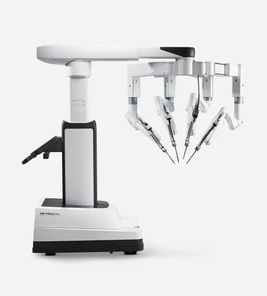 Enlarge ↗
A surgeon-controlled system with a magnified 3D view and wristed instruments, used for procedures including robotic prostatectomy.
Image: Intuitive ↗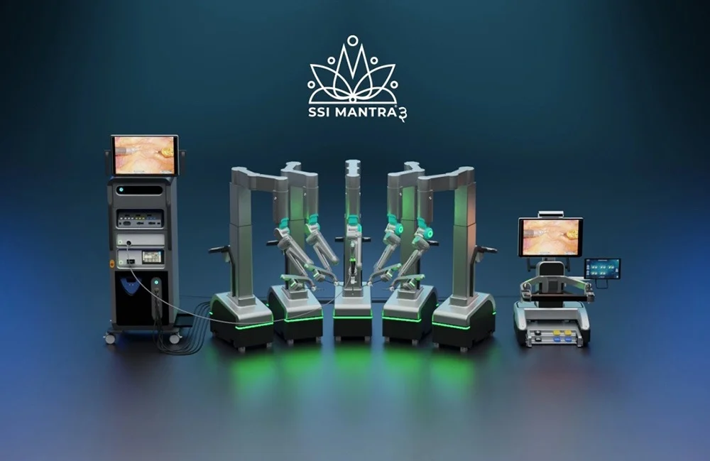 Enlarge ↗
A modular robotic platform with separate instrument arms and a surgeon console. This manufacturer rendering shows the system configuration.
SSI Mantra — India's indigenously developed surgical robotic system. Dr Sachin is among a small group of surgeons experienced on both the da Vinci and SSI Mantra platforms, allowing the right platform to be chosen for each surgery.
Image: SS Innovations via iXBT ↗Different platforms can support minimally invasive surgery. The operation, the surgeon’s experience and individual anatomy matter more than a machine name alone. Explore robotic prostatectomy, Hood and Retzius-sparing approaches ↗
TREATING BENIGN OBSTRUCTION
Lasers can separate, remove or vaporise
obstructing tissue, depending on the technique.
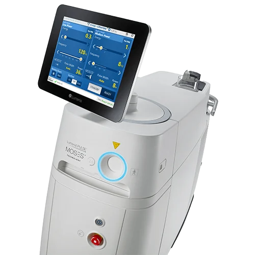 Enlarge ↗
A holmium laser platform. In HoLEP, laser energy separates obstructing prostate tissue before it is removed from the bladder.
Image: Boston Scientific ↗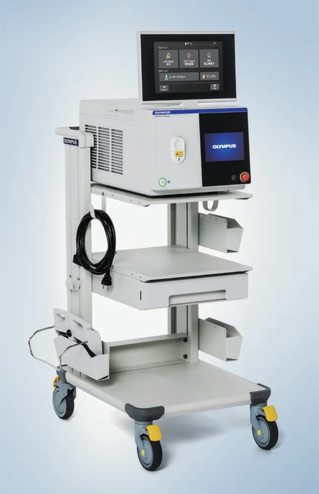 Enlarge ↗
A thulium fibre laser platform for urological surgery. The specific prostate technique and available equipment are discussed with the surgeon.
Image: Olympus ↗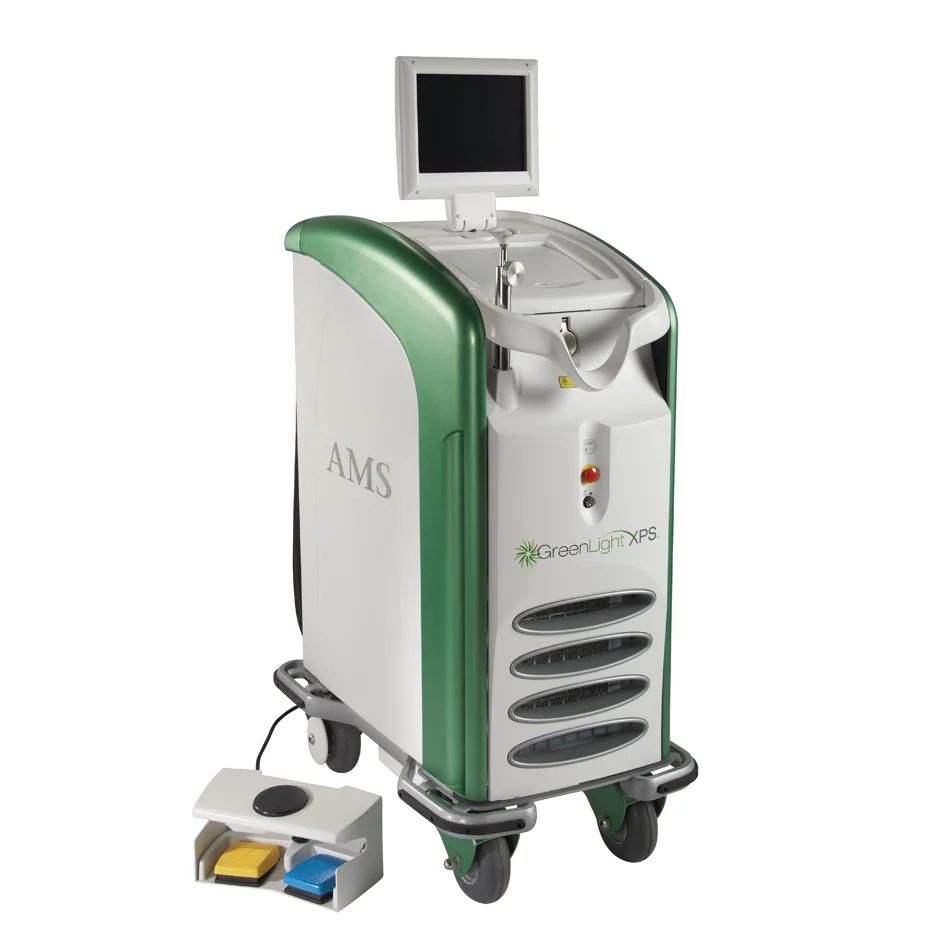 Enlarge ↗
GreenLight laser energy vaporises obstructing prostate tissue to create a more open channel for urine flow.
Image: Boston Scientific ↗HoLEP and ThuLEP separate the enlarged inner tissue, which is then removed.
GreenLight uses laser energy to vaporise obstructing tissue.
TARGETED ULTRASOUND TREATMENT
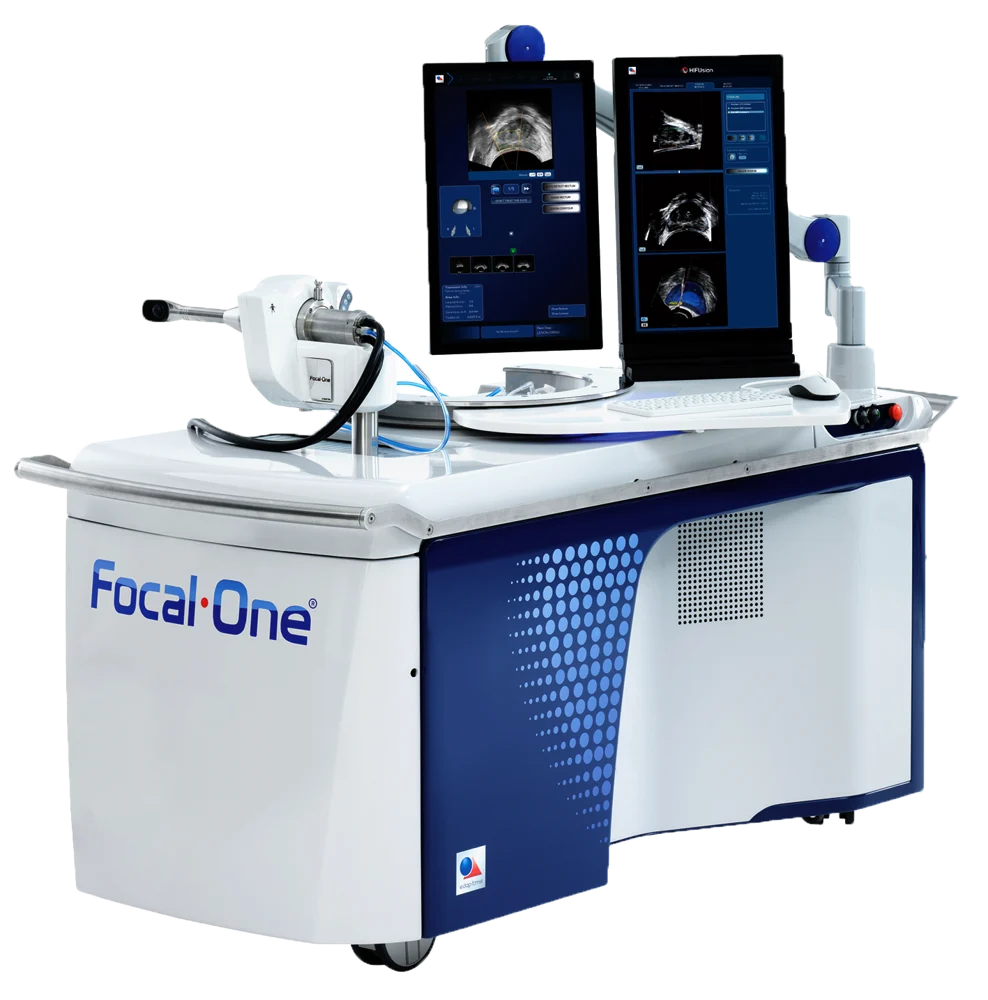
Focused ultrasound energy heats
and destroys selected prostate tissue.
 Enlarge ↗
An image-guided HIFU system that focuses ultrasound energy on selected prostate tissue. Suitability depends on cancer location, extent and individual assessment.
Image: Focal One / EDAP TMS ↗Imaging and biopsy help map the cancer and assess whether focal treatment is appropriate.
Untreated prostate remains. PSA, MRI and repeat biopsy are part of the discussion.
IMAGING GUIDES. TISSUE DIAGNOSES.
A biopsy obtains tissue samples.
The route and targeting method are separate decisions.
 Enlarge ↗
The biopsy needle reaches the prostate through the perineal skin. The ultrasound probe shown in the rectum guides the procedure.
Image: Cancer Research UK / Wikimedia Commons ↗ · CC BY-SA 4.0 · Resized and converted to WebP.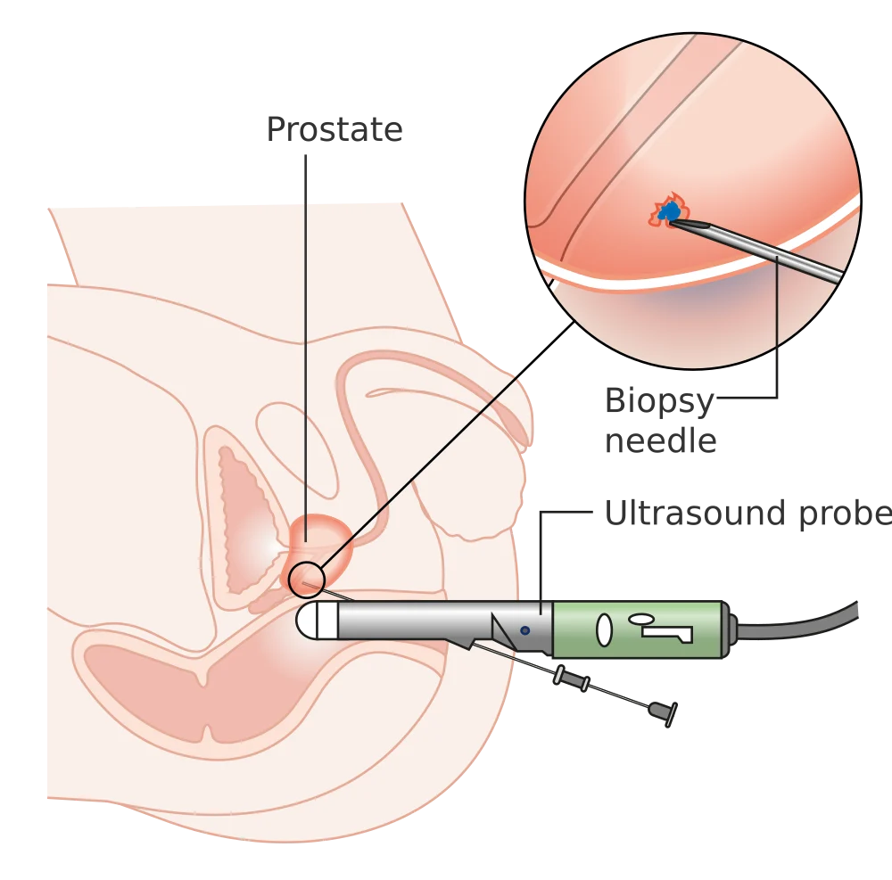 Enlarge ↗
The biopsy needle passes through the rectal wall into the prostate, with ultrasound guidance. The inset illustrates tissue sampling.
Image: Cancer Research UK / Wikimedia Commons ↗ · CC BY-SA 4.0 · Resized and converted to WebP.The needle passes through the skin between the scrotum and anus. An ultrasound probe in the rectum can guide the needle.
The needle passes through the rectal wall. Ultrasound guides sampling. Infection-prevention planning is important.
MRI-visible or micro-ultrasound-visible areas can be sampled. Additional systematic or surrounding samples may be recommended according to the clinical situation.
DIFFERENT TESTS. DIFFERENT QUESTIONS.
Not everyone needs every test.
The clinical question determines the investigation.
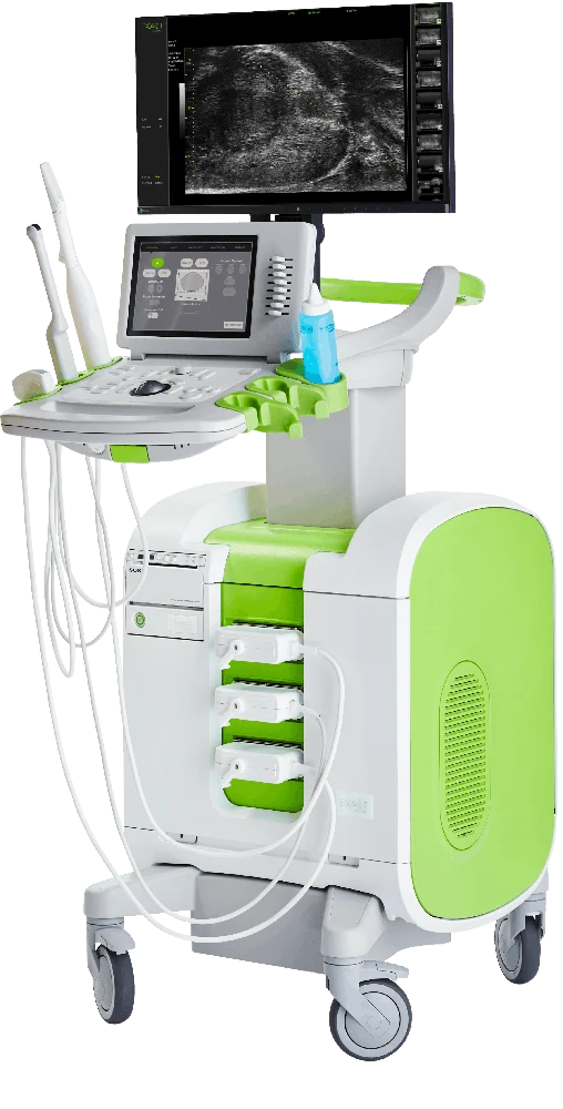 Enlarge ↗
High-frequency ultrasound provides detailed, real-time prostate imaging and can help guide targeted biopsy. Tissue analysis is still needed to diagnose cancer.
Image: Exact Imaging ↗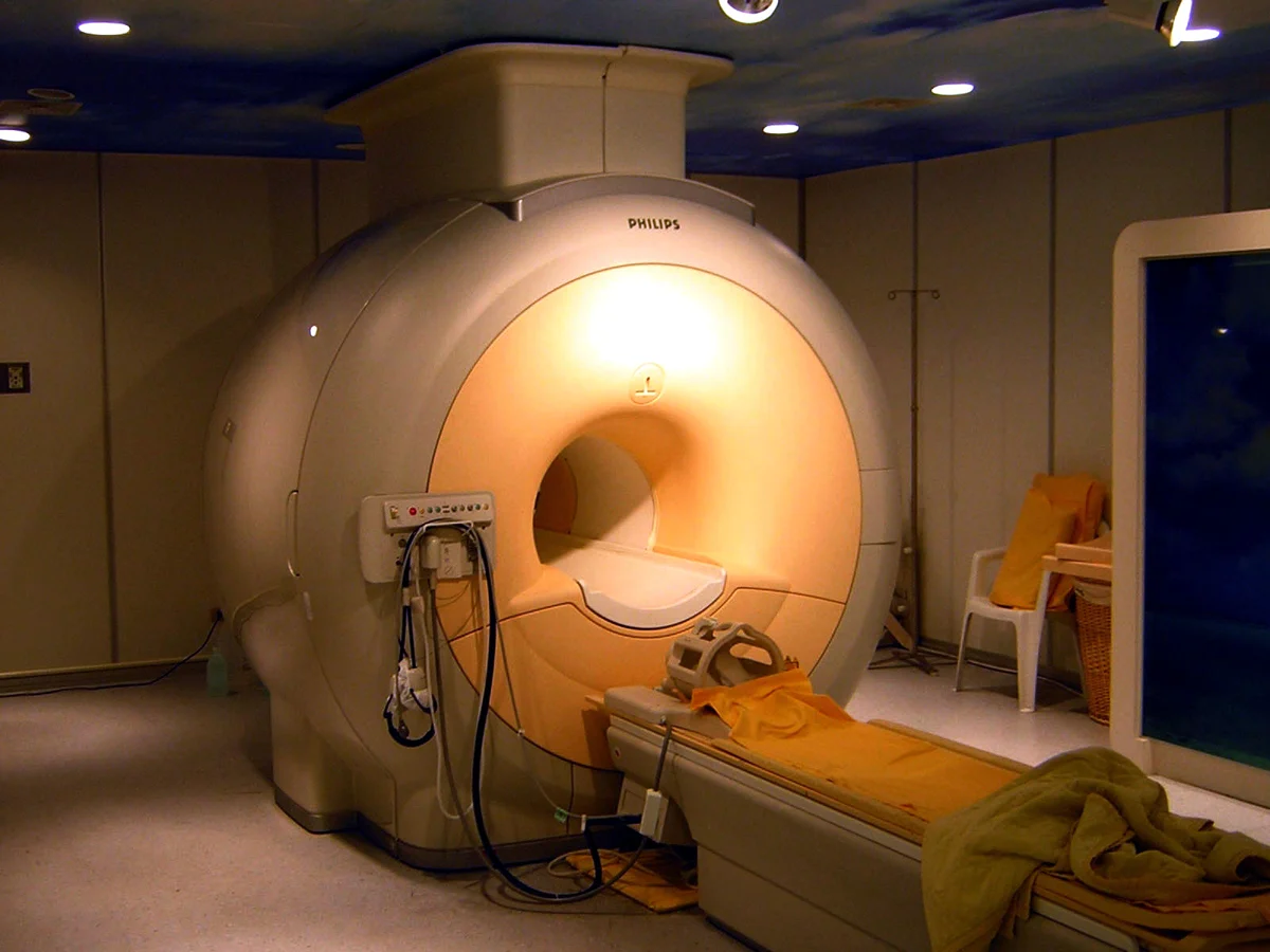 Enlarge ↗
MRI helps assess suspicious areas and local anatomy, often before biopsy. Shown: a Philips Achieva 3T MRI scanner, not a patient scan.
Image: KasugaHuang / Wikimedia Commons ↗ · CC BY-SA 3.0 · Resized and converted to WebP.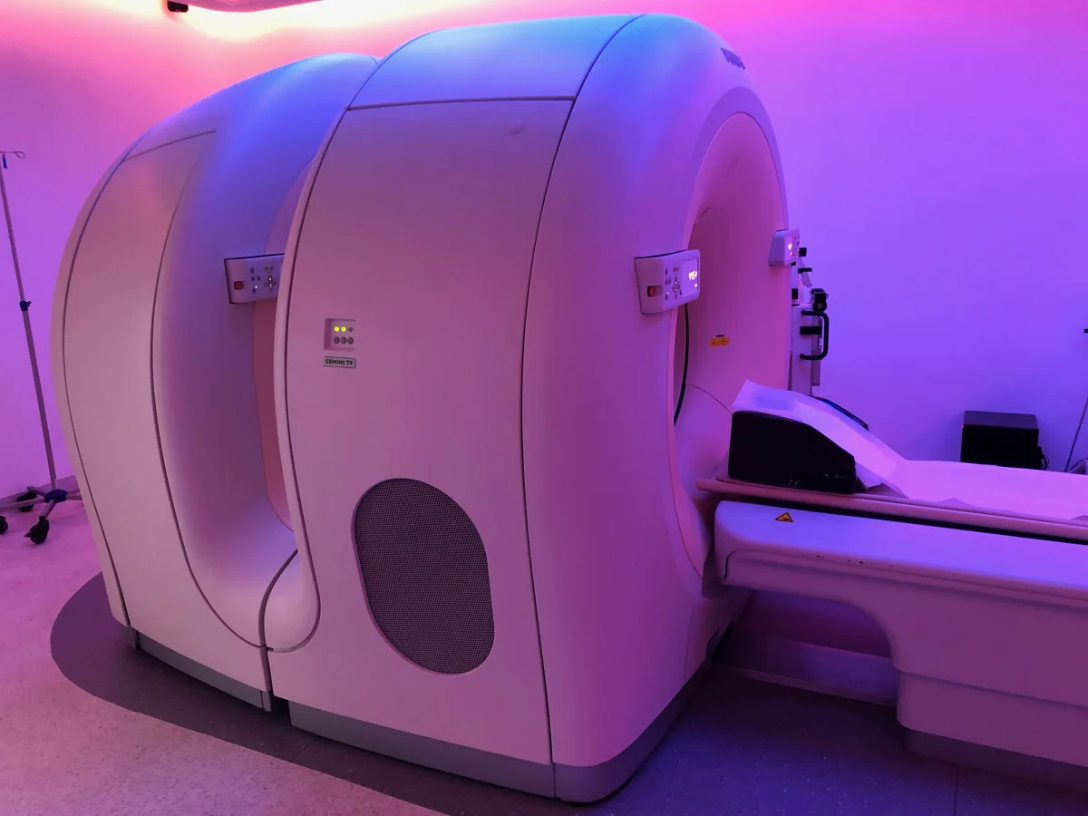 Enlarge ↗
PSMA PET/CT combines a PSMA-targeting radiotracer with PET and CT imaging. Shown: a generic Philips Gemini TF PET/CT scanner; the photograph is not a PSMA scan.
Image: Partynia / Wikimedia Commons ↗ · CC BY-SA 4.0 · Resized and converted to WebP.A PSMA-targeting tracer is given for the scan.
PET shows tracer uptake; CT provides anatomical localisation.
The findings are interpreted with the cancer history and other tests.
PSMA PET/CT can help stage selected prostate cancers or investigate recurrence. It is not a routine screening replacement for PSA, MRI or biopsy. Read the tests and diagnosis guide ↗
PLAN YOUR VISIT
Patients travel from across India and more than 25 countries. Video consultations are available so your reports can be reviewed and a plan agreed before you travel. Max Noida is roughly 40 minutes from Delhi's international airport.
Journey times vary with traffic. Please confirm travel and appointment arrangements with the care team.PATIENT EXPERIENCES
TAKE THE NEXT STEP
Bring your questions, your reports and your priorities.
We’ll discuss the options together.
Sector 128, Noida, Uttar Pradesh
Call +91 76762 73920 ↗OPD: 10 AM – 4 PM, daily · Max Super Speciality Hospital, Sector 128, Noida
Request on WhatsApp ↗Book via Max Healthcare ↗Bengaluru, Karnataka
Bengaluru, Karnataka
For Bengaluru appointments, call +91 76762 73920 or choose a hospital above. Please confirm consultation dates and timings with the care team.